Dr. Ranjan's OrthoExcel Centre
Dr Ranjan Burnwal is extensively trained in treating complex trauma and joint replacement cases.
Book an Appointment
Services
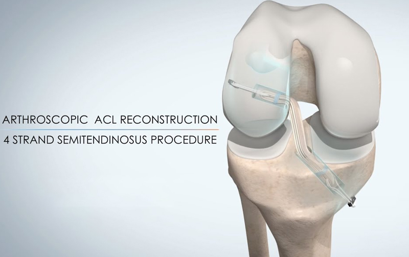
Arthroscopic ACL Reconstruction
Arthroscopic ACL (Anterior Cruciate Ligament) reconstruction is a minimally invasive surgical procedure performed to restore stability and function to a knee with a torn ACL. The ACL is a crucial ligament that connects the thighbone (femur) to the shinbone (tibia) and helps stabilize the knee joint, especially during activities involving sudden stops, pivots, or changes in direction.
ACL injuries are common in athletes and individuals engaged in high-demand sports such as football, basketball, and skiing. When the ACL is torn, it does not heal on its own, and reconstruction becomes necessary, particularly for active individuals or those experiencing knee instability.
The procedure is typically performed arthroscopically, using small incisions through which a camera (arthroscope) and specialized instruments are inserted. This minimally invasive approach reduces tissue damage, promotes faster healing, and allows for better visualization of the joint.
During surgery, the torn ACL is removed and replaced with a graft. The graft may be harvested from the patient’s own body (autograft), commonly using the patellar tendon, hamstring tendon, or quadriceps tendon, or from a donor (allograft). The surgeon drills tunnels into the femur and tibia to anchor the graft in the correct anatomical position. The graft is then secured using screws or other fixation devices.
Post-operative rehabilitation is crucial to restore range of motion, strength, and function. A structured physiotherapy program typically begins within days after surgery and continues for several months. Full recovery and return to sports may take 6 to 9 months, depending on the individual’s progress and adherence to rehabilitation.
Arthroscopic ACL reconstruction is a highly successful procedure that significantly improves knee stability and patient outcomes, allowing individuals to return to their previous level of activity with reduced risk of further injury.